what is a pseudoaneurysm
 Pseudoaneurysm - TeachMeSurgery
Server Error Please try later. Warning: The NCBI website requires JavaScript to operate. NCBI Bookshelf. Service of the National Library of Medicine, National Institutes of Health. StatPearls [Internet]. Treasure Island (FL): StatPearls Publishing; 2021 Jan... StatPearls [Internet]. PseudoaneurysmPhilip A. Rivera; Jeffery B. Dattilo.AuthorsAfiliations Last updated: September 13, 2020. Continuous educational activityThe pseudoaneurismas are false aneurysms that occur on the site of the arterial injury. They are unlike true aneurysms as a layer of the arterial wall does not contain them. Rapid recognition and treatment are required. This activity will cover epidemiology, pathophysiology, evaluation and treatment of pseudoaneurismas most commonly found, including femoral, visceral and aortic pseudoaneurysms. These are separate disease entities that occur in a myriad of situations and require specific training and treatment. This activity reviews the evaluation and treatment of pseudoaneurismas and highlights the role of the interprofessional team in the evaluation and treatment of patients with this condition. Objectives:Introduction An arterial pseudoaneurysm, AKA false aneurysm, is caused by damage to the arterial wall, resulting in locally content hematoma with turbulent blood flow and a neck that normally does not close spontaneously once a certain size has passed. Unlike a true aneurysm, a pseudoaneurisma does not contain any layer of the wall of the glass. On the other hand, there is blood containment for a wall developed with the coagulation cascade products. Eventually, a wall form of fibrin/platelet crosses that is ultimately weaker than those of a true aneurysm. The most common clinical presentation of a pseudoaneurisma is a femoral pseudoaneurysm that follows access to endovascular procedures. Other less common presentations include visceral pseudoaneurismas and aortic pseudoaneurismas. This article will focus on these three entities. Etiology The most common cause of femoral pseudoaneurysm is iatrogenic. The heatrogenic causes include: Visceral artery pseudoaneurysms can occur with catererer-based interventions and are also associated with pancreatitis and pseudocytes. Aortic pseudoaneurysms are often the result of blunt or penetrating trauma, infection, penetrating/degenerative atherosclerotic lesions, and anastomotic sites after vascular bypass or repair. EpidemiologyFemoral pseudoaneurysms often result from access to catheter-based interventions and have an incidence of 0.6 to 4.8%. With the growing use of ultrasound for access, some of the guidelines of society cite that the acceptable rate of pseudoaneurysm after percutaneous access should be less than 0.2 per cent. Visceral pseudoaneurysms are associated with chronic pancreatitis. Pseudoaneurisms of splenic artery were more common but less likely to break. Aortic pseudoaneurysms are often the result of penetrating aortic trauma or injury. Of people with aortic lesions secondary to blunt trauma, 85% die before reaching medical care. 90% of blunt aortic lesions occur only distal to the aortic isthmus and are the result of the deceleration injury of the tethering to arteriosum ligamentum. The aortic pseudoaneurysm is considered a grade III aortic lesion, now more commonly treated with TEVAR instead of open surgery. Rarely, the cause of thoracic aortic pseudoaneurysms is advanced tuberculosis. The research quotes pseudoaneurismas after aortobifemoral bypass at 3.8% in a series of 11 years. PathophysiologyFemoral pseudonyms after percutaneous access may occur due to: Femoral graft anastomosis may also break down over time due to suture failure, or probably more commonly due to the infection of the graft material. Visceral pseudoaneurysms are very rare and are related to the iatrogenic lesion of surgery or endovascular procedures, or pancreatitis, which typically presents as pseudoaneurisma of splenic artery, which is due to the digestive action of pancreatic enzymes in the artery. Unlike the true aneurysms, the visceral pseudoaneurismas appear in countless places. One study found that the distribution of pseudoaneurysms of visceral artery was: celiac axis and branches 39%, liver 39%, splenic 18% and SMA 4%. Aortic pseudoaneurysms due to blunt trauma are theorized to be largely caused by slowing forces between the relatively free aortic arch against the relatively fixed descending aorta, especially at the point where the arteriosum ligamentum anchors the aorta to the pulmonary artery. This fact, in combination with the pinch effect between the breastbone and the spinal column, and the water hammer effect caused by the pressure forces can cause partial rupture of the chest aorta. History and PhysicsTypically, a femoral iatrogenic pseudoaneurysm presents as a painful and pulpit. The dough will often exhibit a brute in the auscultation. If there is an artery-venous fistula, hematoma can demonstrate an a-y-fro quality. The expansion of pseudoaneurysms can exhibit pressure on the skin with pain in the last instance with skin ischemia, necrosis and bleeding. The thrombo formed inside the sack can incarnate in a distual way. Most visceral pseudoaneurysms (91%) have symptoms of rupture. Historically this was called abdominal stroke. Aortic and femoral pseudoaneurysms are seldom spontaneous. Mystical pseudoaneurysms are generally the result of repeated use of drugs IV. Repairing these can be difficult, given the scarcity of veins used for the duct in drug users, as well as the severely infected fields. Evaluation It is necessary to have a complete history and a physical examination. Most cases of femoral pseudoaneurysm are iatrogenic. The results of the physical examination of a pulpit mass in the groin carry a sensitivity of 92% and a specificity of 93% in the diagnosis of a femoral pseudoaneurysm. Duplex ultrasound is still the gold standard for diagnosis, as it can also evaluate for the size, anatomy and the origin of pseudoaneurismas. A CTA helps define the relationship with the surrounding structures, although this test is unnecessary in direct cases. Understanding the characteristics of pseudoaneurysm helps guide treatmentVisceral pseudoaneurysms may present signs of bleeding and abdominal pain. Computed angiography or conventional angiography can help characterize the injury. The diagnosis of aortic pseudoaneurysms is more commonly through conventional CTA or arteryography. They may have a history of prior open repair of dissection or aneurysm, blunt or penetrating traumas, infection or genetic disorders that predispose aorta aneurysm degeneration such as Marfan syndrome or Ehlers-Danlos syndrome. Treatment / ManagementFemoral artery pseudoaneurysms caused by endovascular access used to be treated exclusively by surgical means, but in recent decades, the paradigm has gone to less invasive measures. Management options include observation, ultrasound-guided compression, ultrasound-guided thrombin injection and surgical repair, features of the false aneurysm guide. Femoral artery aneurysms less than 2 to 3 cm in diameter can suffer spontaneous thrombosis and regression with observation. Those who are more rarely thrombos spontaneously, although this is not an absolute rule. Ultrasound-guided thrombin injection is a well-established, minimally invasive method to treat pseudo-aeurismas that are accessible. Several studies have reported a very high success rate (97% to 100%) for femoral pseudoaneurysms with a unique treatment, even while patients are in anticoagulants and/or antiplatelet. The risks of the procedure include embolization and PE, with a shorter aneurysm neck length correlating with a higher risk of ambullic complication, with which they have a neck of less than 2 mm of greater risk. In general, complications are very rare. Ultrasound-guided thrombin injection is not recommended for pseudoaneurysms less than 1 cm due to the theoretical risk of arterial embolization, although these may be safely subjected to an ultrasound-guided compression attempt. The surgical management of the femoral pseudoaneurisma is reserved for those who fail at least a successful compression of duplex or thrombin injection, or for those with anastomotic disorders. If surgical repair is required, the blood must be written and crossed, and available as an inadvertent entry into the pseudoaneurisma before achieving proximal and distal control can result in massive bleeding. The pseudoaneurysms of visceral artery are usually treated by endovascular means first, with surgery reserved for failure; this is very effective, with a study that reports a success rate of 98% in the control of pseudoaneurismas and true broken aneurysms. The techniques include coiling, injections of procoagulating materials and the deployment of covered stent to seal the origin of pseudoaneurisma. Aortic pseudoaneurysms are now treated preferably with TEVAR/EVAR. Even in cases of mycotic pseudoaneurisma and tuberculous pseudoaneurisma of the aorta, stent prosthesis grafts can be saved and avoid the need for a large open operation in already weakened patients. Differential DiagnosisThe differential diagnosis of a femoral pseudoaneurysm includes: PrognosisThe conservative management of femoral pseudoaneurysms has higher rates of failure in the establishment of double antiplatelet therapy, but not for those receiving thrombin injection. Ultrasound-guided thrombin injection for femoral pseudoaneurysms caused by vascular access has a high rate of success (up to 97% to 100%), even in those taking anticoagulants or antiplateletes. Those who fail can undergo a second attempt, and very few must require surgical correction. Although not well studied, endovascular repair of visceral pseudoaneurisma carries a high rate of technical success in small series. Open reparation/ligation is considered more durable, however, and may be a better choice for younger patients. Traumatic aortic pseudoaneurysms treated through TEVAR have an excellent technical success rate (100%) with a low rate of complications related to devices (2.4%) and conversion to open (2.4%). The coverage of the left subclavian artery resulted in a 6% delayed revascularization rate. Complications Femoral pseudoaneurysms can break into retroperitoneal space, causing significant bleeding that may not be immediately obvious, which can lead to death. Complications of ultrasound-guided thrombin injection include distal embolization in up to 2% of patients, although few require any intervention. Complications of endovascular repair of visceral and aortic pseudoaneurysms are mainly related to endovascular devices in general and will not be discussed in depth here. Postoperative and Rehabilitation CareOpen repair of femoral pseudoaneurysms does not require special postoperative care above normal care of vascular patients, and the need for rehabilitation is largely based on the overall condition of the patient prior to the intervention. The endovascular repair of pseudoaneurismas is generally well tolerated, and the patient must remain flat for 2 to 4 hours post-procedure with monitoring for the development of pseudoaneurisma site of subsequent access. Repairs involving the deployment of stent/graft devices will require long-term follow-up with a vascular surgeon for the surveillance image. Consultations For all pseudoaneurysms a vascular consultation of the surgeon is necessary. An interventional vascular surgeon or radiologist may perform ultrasound-guided compression and thrombin injection, but only a vascular surgeon may treat pseudoaneurysms of femoral artery that require surgery. Deterrence and Patient Education There are no specific recommendations for pseudoaneurismas deterrence. The patient must be aware of the signs and symptoms of recurrence. Pearls and Other ProblemsThe moral pseudoaneurysm after vascular access is a common problem and can be addressed with less invasive means than open surgery. Pseudoaneurismas that occur in the mandate of vascular anastomosis repair and open review. Visceral pseudoaneurysms are rare, and the diagnosis of these is often during an investigation for vague symptoms of abdominal pain, or with CTA images with signs of bleeding. Aortic pseudoaneurysms are rarely spontaneous and result from infection or trauma. Improvement of Health Team Results The management of pseudoaneurysms requires an interprofessional team approach, including doctors, specialists and specialized nurses who collaborate in disciplines to achieve optimal patient outcomes. The management of pseudoaneurisma depends on its etiology and location. Nurses and doctors should routinely palpate the English area after patients undergo cardiac catheterization, as pseudoaneurysms are not rare. The vascular surgeon may treat a femoral pseudoaneurysm or interventional radiology with thrombin injection depending on local practice patterns. Endovascular repair of pseudoaneurysms of visceral artery can be performed by a vascular surgeon or interventional radiologist as well. Aortic pseudoaneurysms are typically the domain of the vascular surgeon as there is a possible need for open repair and subsequent follow-up. Only the vascular surgeon can perform open repairs of all types of pseudoaneurismas. [Level V] Continuous Questions of Education / Examination Left ventricular pseudonym. Image courtesy S Bhimji MD References This book is distributed under the terms of the Creative Commons 4.0 International License (), which allows the use, duplication, adaptation, distribution and reproduction in any medium or format, provided that you give appropriate credit to the original author(s) and source, a link to the Creative Commons license is provided, and changes are indicated. ViewsIn this PageRelated information Similar products in PubMed Recent activityYour navigation activity is empty. The activity recording is off. , 8600 Rockville Pike, Bethesda MD, 20894 USA

Contemporary Management of Postcatheterization Pseudoaneurysms | Circulation

Pseudoaneurysm - TeachMeSurgery

Pseudoaneurysm - Wikipedia

Incidence and predictors of post-catheterization femoral artery pseudoaneurysms - ScienceDirect

False aneurysm | Radiology Reference Article | Radiopaedia.org

Surgical treatment of a pseudoaneurysm of the femoral artery secondary to a gunshot wound. Clinical case report | Medicina Universitaria

Pseudoaneurysm - an overview | ScienceDirect Topics

Pseudoaneurysm - TeachMeSurgery
 | Stanford Health Care")
Types of Thoracic Aortic Aneurysm (TAA) | Stanford Health Care

Femoral site psudeoaneurysm

Schematic diagram of differences between true aneurysm and... | Download Scientific Diagram

False aneurysm | Radiology Reference Article | Radiopaedia.org
Preservation of the superficial temporal artery during resection of a pseudoaneurysm: A case report and review of the literature

Pseudoaneurysm of Neck External Carotid Artery Related to Chiropractic Manipulation

Endovascular Treatment of a Femoral Pseudo-aneurysm - YouTube

Aneurysm | Johns Hopkins Medicine

Spontaneous Pancreaticoduodenal Artery Pseudoaneurysm Rupture

86- Abdominal Aortic Aneurysm Flashcards | Quizlet

Iatrogenic femoral artery pseudoaneurysm surgically repaired with combined bovine pericardial roll and autologous great saphenous vein grafts - ScienceDirect

Femoral pseudoaneurysms after percutaneous access - Journal of Vascular Surgery

Treatment of Iatrogenic Pseudoaneurysm of the Brachial Artery: Case Report and Literature Review | Vascular Disease Management

Case of the week - superficial femoral artery pseudoaneurysm

$3M VERDICT IN FAILED CARDIAC PROCEDURE | ADM

Pseudoaneurysms and the Role of Minimally Invasive Techniques in Their Management | RadioGraphics

Transforming Growth Factor-β1 Inhibits Pseudoaneurysm Formation After Aortic Patch Angioplasty | Arteriosclerosis, Thrombosis, and Vascular Biology

Iatrogenic Pseudoaneurysms | IntechOpen

Fluoroquinolones Side Effects Lawsuit | Beasley Allen Law Firm

Aneurysm - AMBOSS

Cureus | Superficial Femoral Artery Pseudoaneurysm and Arterial Wall Destruction After Drug-Coated Balloon Treatment

Treatment of Post-Catheterization Femoral Artery Pseudo-Aneurysm With Para-Aneurysmal Saline Injection - American Journal of Cardiology

Femoral pseudoaneurysms after percutaneous access - ScienceDirect

Pseudoaneurysms and the Role of Minimally Invasive Techniques in Their Management | RadioGraphics
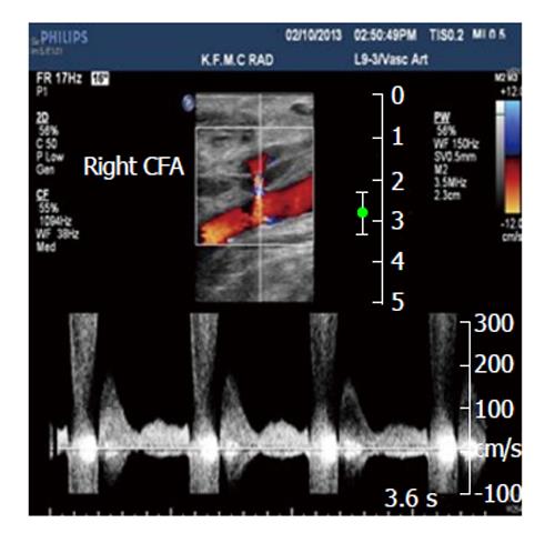
To-and-fro" waveform in the diagnosis of arterial pseudoaneurysms

Pseudoaneurysm | Cedars-Sinai

Iatrogenic femoral artery pseudoaneurysm surgically repaired with combined bovine pericardial roll and autologous great saphenous vein grafts - ScienceDirect

Common femoral artery pseudoaneurysm | Radiology Case | Radiopaedia.org

Treatment of Iatrogenic Pseudoaneurysm of the Brachial Artery: Case Report and Literature Review | Vascular Disease Management

Case of the week - superficial femoral artery pseudoaneurysm

What is PSEUDOANEURYSM? What does PSEUDOANEURYSM mean? PSEUDOANEURYSM meaning & explanation - YouTube

Duplex Ultrasound: Evaluation and Management of Post-catheterization Femoral Pseudoaneurysms | SpringerLink
Posting Komentar untuk "what is a pseudoaneurysm"